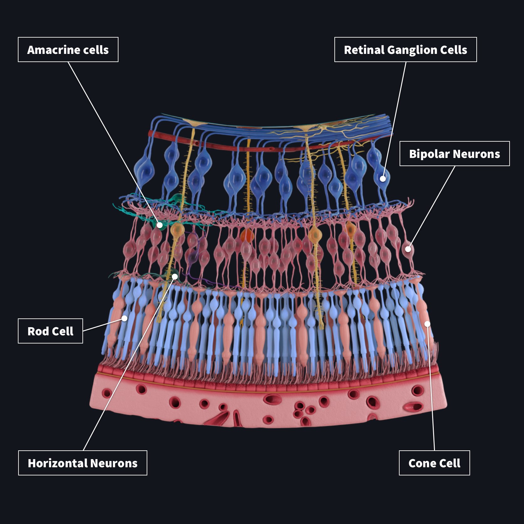

Heidelberg Engineering's growing product portfolio combines these core technologies: confocal microscopy, scanning lasers and optics, optical coherence tomography (OCT), real-time image processing and analytics, multimodal image management solutions (PACS), electronic medical records (EMR) and large-scale data analysis. Retinal Layers in 1 minute / Mnemonic series 13/Retinal layers mnemonic/Retinal layers anatomy /Retinal layers diagram. The company's substantial expertise in the development and implementation of intelligent image and data management solutions complements its distinguished history in the design, manufacture and distribution of ophthalmic diagnostic instruments. Uncompromising quality and education play a large part in fostering the diagnostic confidence that has become synonymous with the global brand. tomography (OCT) allows visualization of the vitreous, retinal layers, retinal pigment. From its inception in 1990, the company has collaborated with scientists, clinicians and industry to develop innovative products that deliver clinically relevant benefits. The vitreous, retinal layers and choroidal layers are visible. The retinal nerve fiber layer and retinal ganglion cell layer are affected in conditions like glaucoma. Certain types of retinal conditions can affect different retinal layers. OCT progression analysis technology has improved greatly.

OCT’s need to overlay circum presentations taken over time with actual NFL thickness to allow improved comparison, not quadrant colors. Heidelberg Engineering continuously optimizes imaging and healthcare IT technologies to provide ophthalmic diagnostic solutions that empower clinicians to improve patient care. Each distinct retinal layer is visible on the OCT and corresponds well to histological studies. Glaucoma diagnostic ability of layer-by-layer segmented ganglion cell complex by spectral domain optical coherence tomography.


 0 kommentar(er)
